Imaging and characterisation
Our researchers
- Timothy Burnett
- Robert Cernik
- Philip Edmondson
- Alexander Eggeman
- Ali Gholinia
- Sarah Haigh
- Wajira Mirihanage
- Katie Moore
- Andrew Thomas
- Philip Withers
Our Department has one of the most extensive and innovative imaging and characterisation facilities in the UK - something that our researchers are able to take advantage of in their work.
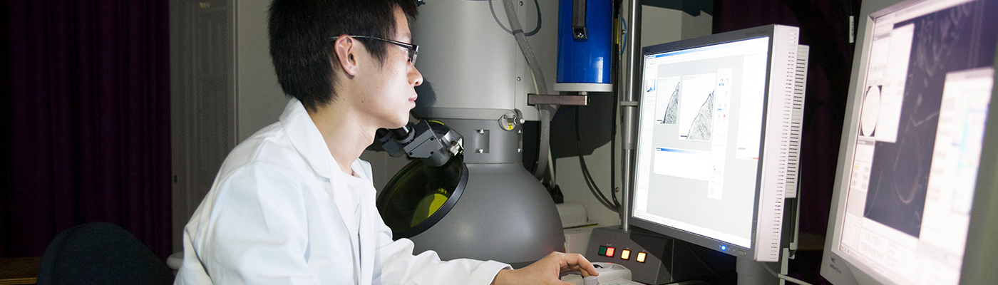
By using a suite of tools that includes modern X-ray and microscopy facilities, we're able to gain a better understanding of advanced materials.
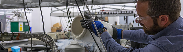
In-situ characterisation allows us to study the performance of materials in demanding environments (for example in hydrogen gas, or at temperatures up to 1,100 °C).
Our imaging and characterisation research brings together world-renowned academics in Manchester, making use of the extensive facilities available.
Case studies
Imaging and characterisation research is making a significant societal impact, as demonstrated by our case studies:
Correlative Imaging
Manchester has a vast range of characterisation facilities allowing imaging and analysis at length scales spanning from atoms to metres and time scales from nanoseconds to weeks. Our researchers have a combined interest in correlative imaging where several different characterisation approaches are combined on the same specimen, often on the same specific area, in order to gain a deeper understanding of a particular materials phenomena.
For example, a study has been performed in collaboration with BP to shed light on the mechanism of corrosion for a steel pipeline; starting from the full component and digging down to atoms at a crack tip. Most recently tomographic imaging and elemental analysis capabilities are being used to extend correlative imaging to three-dimensional structure and chemistry.
Uncovering atomic defects in 2D Material Electronics
Working closely with colleagues in the National Graphene Institute we have been able to delve into the structure of 2D materials at the atomic scale using our Titan ChemiSTEM transmission electron microscope. By imaging stacks formed of several different 2D materials we have shown that some are air sensitive, degrading over time - even in air.
We have shown that the resulting atomic defects will prevent their exciting properties being exploited unless these can be removed by processing in an inert atmosphere. We have also imaged the structure of graphene nanocapillaries, which has helped to understand unusually fast flow rates observed when structures are just a few molecules wide.
Tomography keeps its cool to analyse ice cream
Using 3D in situ X-ray tomography (XRT) it is possible to visualise the effects of changing temperature on the microstructure of ice cream. Ice cream contains milk, fats, sugars, proteins, emulsifiers, stabilisers and flavours that are aerated and then frozen. Its quality depends on the microstructure (grain size of ice crystals and porosity): smaller crystals and bubbles make it smoother and creamier. This complex colloid is unstable above -30˚C so its microstructure will change during shipping and storage (domestic freezers are usually at around -18˚C), which will affect its taste and texture. We have used X-ray CT to analyse the structure of ice cream to help develop routes to keep it tastier for longer.